Triangular Fibrocartilage Complex

Lower Back Pain
August 10, 2020
Tennis Elbow
August 27, 2020Triangular Fibrocartilage Complex

Triangular Fibrocartilage Complex Injury (TFCC)
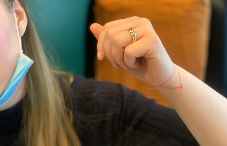
The triangular fibrocartilage complex (TFCC) is a load-bearing structure that sits on the pinky side of the palm between the wrist and the hand- or more specifically between the lunate, triquetrum, and ulnar head.
It’s a very complicated structure that spans a large number of joints and is comprised of many different structures (read the rest of this paragraph only if you want the down-and-dirty anatomical specifics!). It includes the triangular fibrocartilage disc, extensor carpi ulnaris tendon subsheath, ulnotriquetral and ulnolunate ligaments, meniscal homolog, dorsal and volar distal radioulnar ligaments, and the ulnocarpal collateral ligament. The triangular fibrocartilage disc attachment on the radial side (thumb side) is to hyaline cartilage, which makes this weaker compared to the bony attachment on the ulnar side’s (pinky side). [Casadei & Kiel 2019]
Because the TFCC is so broad in its coverage and jobs, this can mean that injuries and symptoms can be highly variable and consequently treatment can range from a week or 2 of soreness to even surgery to restabilise the wrist. In short, your rehab plan will vary significantly based on blood supply alone- parts of the TFCC are well vascularised giving them a decent ability to regenerate and heal whereas other segments have no blood supply at all. The outside of the TFCC (between 10 and 40% of it) is well vascularised giving it a fairly decent ability to heal. The central portion, however, is avascular meaning it has no blood supply. Additionally, the fibrocartilage on the ulnar side (pinky side) is vascularised but the rest is not.
Most commonly we see a TFCC injury with a fall onto an outstretched hand landing on a palm down (pronated and extended), or from a forceful rotation or distraction. As the TFCC is a stabiliser of the distal radioulnar joint (DRUJ) and ulnar side of the wrist any forces that place a stress on these areas will tend to cause TFCC injuries. Sports with racquets or bats can also place stress on the TFCC due to the repeated ulnar deviation.
Symptoms are usually very localised to the pinky side of the wrist, pain that increases when the wrist is moved from side to side, painful clicking of the wrist and pain with or loss of grip strength (opening a jar, lifting a fry pan or oven tray).
Treatment is highly variable and mainly comes down to the specific grade of the injury. If you’ve injured a part of the TFCC that is responsible for stability, then we may place you into a specific splint for a period of time to allow the ligaments to heal up. The Internet will tell you to completely immobilise the wrist but often this is overkill for the majority of injuries we see in the clinic.
Treatment for the majority of TFCC injuries is usually 2-6 weeks’ worth and consists of unloading with bracing or tape, followed by a progressive and specific strengthening program to rebalance and restabilise the wrist.
If you want to know the specifics on the TFCC grading I have included it below. It is very dry and anatomical but really outlines why a consult is needed to determine specifically the path you need to take to get a good outcome!
There are actually 2 classes of injury to the TFCC which basically come down to
Class 1: traumatic
Class 2: degenerative
Within the 2 classes there are subtypes which narrow down specifically what was injured, where and to what extent.
1A: The central aspect of the triangular fibrocartilage disc is perforated. Since the radioulnar ligaments are intact, these are stable injuries.
This injury is in an avascular region that will not heal if it does not receive treatment. Due to the lack of vascularity, it does not respond to direct surgical management, so debridement is the intervention of choice
1B: Injury of the ulnar attachment of the triangular fibrocartilage disc.
The area has vascularization, so direct surgical repair is an option. If the triangular fibrocartilage disc is completely detached from the ulnar insertion, then there is an injury to the radioulnar ligaments, and there will be instability. If this is the case, the amount of retraction of the tendon should be measured, and a tendon graft may be necessary as part of the surgical repair. Partial tears would not involve radioulnar ligament injury and thus are stable and could be treated with sutures arthroscopically. Tears at the foveal insertion require bony reattachment, and therefore these are of more significant consequence than styloid insertion tears
1C: Distal disruption of the TFCC. In this case, there is a detachment of the volar ulnotriquetral and ulnolunate ligaments from the carpal attachment.
Arthroscopy and debridement are both options.
1D: Injury to the radial attachment of the triangular fibrocartilage disc. These injuries are communicating on MR arthrography.
If the injury involves radioulnar ligament damage, surgical reattachment is the treatment of choice. If the injury spares the radioulnar ligaments, partial resection via arthroscopy in an option
Type 2 is a more degenerative type injury and so the grading is more focused around the level and extent of wear. The treatment is separated by whether the lunotriquetral ligament is torn or intact. Type 2A, 2B, and 2C lesions often do well with conservative therapy such as splinting and retraining with physio. Type 2E and 2D lesions are often treated surgically.
For patients with chronic tears who undergo surgery, one study of 57 patients who had pain for nine months on average prior to surgery found a 98% satisfaction and return to work around nine weeks. [Mathoulin 2017]
2A: Degenerative changes of the triangular fibrocartilage disc without evidence of perforation.
2B: Grade 2A with the additional presence of chondromalacia of the hyaline cartilage on the articular surface.
2C: Full thickness perforation of the triangular fibrocartilage disc.
2D: Any of the features in 2A through 2C plus lunotriquetral ligament tear.
2E: Grade 2D with the additional presence of ulnocarpal arthritis.

