City Physio & Pilates | Expert Insights | Low Back Pain
It Feels Like Sciatica. It Might Not Be.
Shooting leg pain is one of the most misattributed symptoms in musculoskeletal practice. Here’s how to tell whether the problem is in your spine — or your backside.
The pain starts in the buttock. It travels down the back of the thigh, sometimes into the calf, occasionally into the foot. It burns, or aches, or produces a pins-and-needles sensation that is difficult to describe and even harder to ignore. Sitting makes it worse. Your GP says sciatica. Everyone you mention it to says sciatica. The internet says sciatica. ChatGPT says sciatica…
Sciatica is a symptom, not a diagnosis. It describes irritation of the sciatic nerve, the longest nerve in the body, which produces exactly the pattern above. What it does not tell you is where the nerve is being irritated, or why. And that distinction is the entire ballgame when it comes to treatment.
Two of the most common causes of sciatic nerve irritation are lumbar disc pathology (what most people mean when they say sciatica) and piriformis syndrome – compression of the sciatic nerve at the level of the hip, by the piriformis muscle. (Uhhh I hate the name piriformis syndrome! But I’ll get to that!)
Piriformis Syndrome produces nearly identical symptoms to sciatics. They require quite different management. Getting one confused for the other is exceedingly common and explains a lot of prolonged, treatment-resistant leg pain.
… OK, let me explain my hatred of the name Piriformis Syndrome.
The piriformis is a muscle within the pelvis. It rarely becomes tight for no reason. In fact, it can become tight for a variety of reasons which SHOULD be the name for the condition, not just naming a muscle. Most commonly, a pelvic instability or asymmetry can cause it to spasm or tighten giving the classic “piriformis syndrome” diagnosis. But, (and here comes the bit where I hate it the most) if we just look at the piriformis it is an incomplete picture and a recipe for recurrence! If you find out why it is tight or spasming and correct that, then the muscle should relax and you can then layer in from there. The piriformis is the hot spot, not the “cause”.
The anatomy in brief
The sciatic nerve is formed from nerve roots L4 to S3, which exit the lumbar and sacral spine and converge into a single nerve that travels through the pelvis, exits beneath (and occasionally through) the piriformis muscle, passes through the greater sciatic foramen, and runs down the posterior thigh before dividing into the tibial and common peroneal nerves near the knee.
It can be compressed or irritated at any point along this path. The two most clinically significant points of compression are:
- At the intervertebral foramen — where a disc bulge or herniation, degenerative bony change, or foraminal stenosis compresses the exiting nerve root before it joins the sciatic nerve. This is true radiculopathy.
- At the piriformis muscle — where a hypertrophied, spasmed, or anatomically variant piriformis compresses the sciatic nerve in the deep gluteal space. This is piriformis syndrome, also called deep gluteal syndrome.
True sciatica from lumbar pathology: what it looks like
Lumbar radiculopathy, nerve root compression in the spine, produces a specific, dermatomal pattern of symptoms. The distribution of pain, numbness, and weakness corresponds to the nerve root affected:
- L4: Inner calf and medial foot; weakness in knee extension
- L5: Outer calf, top of foot and big toe; weakness in big toe and foot extension (foot drop in severe cases)
- S1: Outer foot and small toes; weakness in plantarflexion; reduced or absent Achilles reflex
Features that support a lumbar origin include pain that worsens with lumbar flexion (sitting, bending forward) or extension depending on the mechanism, a positive straight leg raise test at less than 60 degrees, neurological signs such as reflex changes or dermatomal sensory loss, and a clear association with a precipitating event such as a heavy lift or a period of sustained lumbar loading.
Piriformis syndrome: what makes it different
Piriformis syndrome produces sciatic nerve symptoms- buttock pain radiating into the posterior thigh, sometimes the calf- without any lumbar spine involvement. The nerve is being compressed peripherally, not centrally, which changes the clinical picture in several important ways.
Features that suggest piriformis syndrome
- Buttock pain that is provoked by direct pressure over the piriformis muscle belly – deep in the gluteal region, not the lumbar spine
- Pain that worsens with prolonged sitting, particularly on hard surfaces or with the hip in flexion and internal rotation (crossing the legs, sitting in low chairs)
- Reproduction of symptoms with specific hip movements – particularly resisted external rotation or passive internal rotation of the hip in flexion
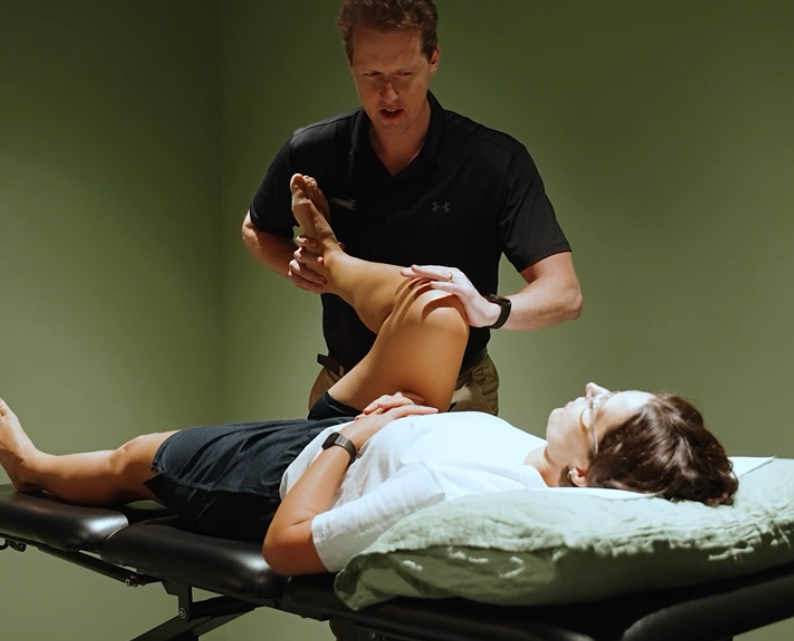
- A positive FAIR test (Flexion, Adduction, Internal Rotation) – the clinical provocation test most associated with piriformis syndrome
- Absence of neurological signs – no reflex changes, no dermatomal sensory loss, no weakness in the distribution of a specific nerve root
- Negative or equivocal straight leg raise
- No relevant findings on lumbar spine imaging – a lumbar MRI that is normal or non-contributory in someone with leg pain is a significant pointer toward a peripheral source
Why piriformis syndrome gets missed
Several reasons. First, the symptom pattern is genuinely similar – posterior leg pain is posterior leg pain, and without a careful clinical examination most presentations get labelled sciatica and treated as lumbar in origin. Second, imaging tends to look at the spine. A lumbar MRI is the standard investigation for leg pain; it doesn’t image the piriformis or the sciatic nerve at the hip unless someone specifically requests it. Third, some degree of disc degeneration is almost universal in adults over 40, so lumbar MRI findings are easy to find and easy to blame – whether or not they’re actually producing the symptoms.
The result is that patients with piriformis syndrome are frequently treated with lumbar-focused physiotherapy, lumbar epidural injections, or – in the worst cases – lumbar spinal surgery that does nothing to resolve symptoms, because the spine was never the problem.
The treating clinician also needs to assess and adequately address the reasons that could be causing the piriformis to grip, including subtle pelvic instabilities, pelvic asymmetry, postural habits, overactive hip flexors and trunk rotations/ contributions
Treatment: where the two approaches diverge
For lumbar radiculopathy
Management is directed at the spine: neural mobilisation, lumbar joint and disc offloading, specific directional exercise (typically extension-biased for posterolateral disc herniations), progressive lumbar stabilisation, and in some cases referral for imaging-guided injection or surgical opinion for significant neurological deficit.
For piriformis syndrome
Management is directed at the hip, SIJ, pelvis and deep gluteal region: piriformis and deep external rotator stretching and soft tissue release, sciatic nerve neural mobilisation from the peripheral end, hip external rotator strengthening in functional ranges, and addressing the underlying hip control and loading patterns that drove the piriformis into overactivity in the first place. For the pelvis we look at stability and control, subtle changes through the thorax and hip joints to improve the system wholistically.
At City Physio & Pilates, assessment includes a thorough examination of the body as a whole to determine where the nerve is being loaded – and treatment is directed accordingly. In some cases, both structures contribute and need to be addressed in sequence.
Frequently asked questions
Both produce sciatic nerve pain — buttock and leg pain that may extend to the calf or foot. True sciatica originates from nerve root compression in the lumbar spine, typically from a disc herniation or bony stenosis. Piriformis syndrome originates from compression of the sciatic nerve at the hip, by the piriformis muscle. Same pain. Different source. Different treatment.
Clinically… through history and physical examination. Key findings include deep gluteal tenderness over the piriformis, pain provoked by the FAIR test (hip flexion, adduction and internal rotation), absence of neurological signs such as reflex changes or foot weakness, and a lumbar spine that doesn’t reproduce the leg symptoms on examination.
No. A standard lumbar MRI doesn’t image the piriformis or the sciatic nerve at the hip. Additionally, an MRI doesnt pick up tension or dynamic instability- these can only be seen with a specific clinical assessment. A normal lumbar MRI in someone with leg pain is actually useful information, it suggests the spine isn’t the problem, and a peripheral source like piriformis syndrome should be on the list.
AND that is why we’re here! Yes, and it generally responds well. Treatment includes soft tissue release of the deep hip rotators, sciatic nerve mobilisation, hip strengthening, and correcting the movement patterns that drove the piriformis into overactivity in the first place.