City Physio & Pilates | Knee Pain | Martin Place, Sydney CBD
Your Knee Hurts. But Is Your Knee Actually the Problem?
The hip is one of the most prolific sources of referred pain in the lower limb… and one of the most reliably overlooked. Here’s why knee pain isn’t always a knee story.
You’ve had knee pain for four months. You’ve had it imaged. The MRI showed some mild wear, possibly a small meniscal change, nothing surgical. You’ve done six weeks of knee-focused physiotherapy you found on ChatGPT; VMO strengthening, patellar taping, step-ups. It hasn’t helped much. The knee still aches on the stairs, still nags after a long day on your feet, still wakes you occasionally when you roll onto that side at night.
Here is a question nobody has asked you yet: has anyone examined your hip? More specifically, your lumbopelvic control?
The hip is one of the most common sources of referred pain to the knee, and one of the most consistently missed in clinical practice. Not because it’s subtle, a thorough hip examination takes about five minutes, but because pain located at the knee creates a powerful cognitive pull toward the knee as the source. The patient points to their knee. The clinician looks at the knee. The hip, sitting quietly a little north continues doing exactly what it’s been doing all along.
Why the hip refers pain to the knee
Referred pain from the hip to the knee is not a curiosity, it’s an anatomical inevitability. The obturator nerve, which provides sensory innervation to the hip joint, also sends branches to the medial knee (the outside of the knee). The femoral nerve and its cutaneous branches supply both the anterior hip and the anterior thigh down toward the knee. When the hip joint is under sufficient mechanical or inflammatory stress, pain signals travel these shared pathways and are interpreted by the brain as originating in the knee.
This is the same mechanism behind referred pain anywhere in the body, the cervical spine producing headaches, the thoracic spine producing rib pain, the lumbar spine producing leg pain. The brain maps pain to where it expects the signal to come from based on shared neural territory, not necessarily where the problem actually is.
Hip osteoarthritis is the most common culprit, but it isn’t the only one. Femoroacetabular impingement (FAI), labral tears, hip flexor tendinopathy, and even referred pain from the lumbar spine via the L3 nerve root can all produce anterior knee or medial knee symptoms without any local knee pathology to explain them. Additionally, a hip and pelvis that is not producing adequate stability with load transfer (running, stairs etc) causes lateral shift of the body relative to the knee. This *can* cause knee pain, but it’s certainly just the symptom and not the cause!
The clinical features that point toward the hip
Several patterns, taken together, should raise strong suspicion that the hip rather than the knee is driving the symptoms.
Pain location and behaviour
- Anterior knee or medial knee pain without a clear local mechanism- no injury, no specific provocative activity at the knee itself
- Pain that is diffuse and difficult to localise precisely, rather than pointing to a specific structure
- Groin pain or anterior thigh aching accompanying the knee symptoms- often reported as a separate complaint rather than connected to the knee
- Pain that is worse with prolonged walking, stairs, or getting up from a low chair- activities that load the hip as much as the knee
- Night pain, particularly on the affected side when lying with the hip in adduction and internal rotation
What the examination finds
- A knee examination that is largely unremarkable; no joint line tenderness, no meniscal provocation signs, no ligamentous laxity, no effusion
- Restricted hip range of motion, particularly internal rotation and flexion; the most sensitive examination finding in hip osteoarthritis
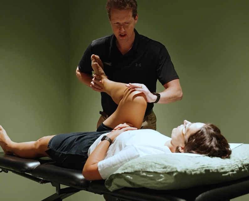
- Pain reproduced at the groin or anterior thigh with hip provocation testing; the FADIR test (flexion, adduction, internal rotation) or the FABER test (flexion, abduction, external rotation), rather than at the knee
- Antalgic gait or a subtle Trendelenburg pattern; the pelvis drops on the contralateral side during single-leg stance, indicating hip abductor weakness or inhibition. This is also observed with single leg hopping, single leg squats or step ups.
- Imaging of the knee that is normal or shows only minor age-related changes inconsistent with the symptom severity
Hip osteoarthritis: the most commonly missed source
Hip osteoarthritis (OA) deserves particular attention here because it is both extremely common and extremely good at hiding. Classic hip OA presents with groin pain and restricted rotation- but a significant proportion of patients present with anterior thigh or knee pain (and sometimes back pain!) as the dominant complaint, with no groin symptoms at all. Studies suggest that up to 45 percent of patients with confirmed hip OA report knee pain as part of their presentation, and a meaningful subset report knee pain as their primary complaint.
The clinical picture that should trigger hip examination: a patient over 45 with knee pain of gradual onset, mild morning stiffness, and no clear mechanism of injury, whose knee examination is underwhelming relative to the symptom severity. Add restricted hip internal rotation on examination and the diagnosis becomes very probable before imaging is even ordered.
X-ray of the hip, standing, weight-bearing, is the appropriate first imaging investigation when hip OA is suspected. Not the knee.
Femoroacetabular impingement and labral pathology in younger patients
In patients under 45, the hip pathologies most likely to refer to the knee are femoroacetabular impingement and labral tears. FAI is a morphological condition in which abnormal bony contact between the femoral head and the acetabular rim occurs during hip flexion; producing pain in the groin, anterior thigh, and occasionally the knee, particularly with sitting, squatting, and rotational activities.
These patients are frequently active, often presenting after an increase in training load or a change in activity. Their knee examination is clean. Their hip examination, particularly FADIR provocation, is not. They’ve often been through a cycle of knee-focused treatment before anyone thinks to look at the hip, partly because FAI is underdiagnosed generally, and partly because the knee pain is the louder complaint.
True Knee Pathology
- Pain localises precisely to knee structures
- Clear mechanism- injury, overload, sport
- Positive meniscal or ligament tests
- Joint line tenderness on palpation
- Effusion or swelling at the knee
- Imaging consistent with symptoms
- Hip examination largely normal
Referred Pain from Hip
- Diffuse anterior or medial knee pain
- Gradual onset, no clear knee mechanism
- Knee examination underwhelming
- Restricted hip IR and flexion
- Positive FADIR or FABER test
- Groin or anterior thigh symptoms
- Poor response to knee-directed treatment
What happens when you treat the right joint
The good news, and it genuinely is good news, is that when the hip is identified as the source, knee symptoms typically respond well to hip-directed treatment. Hip OA management includes progressive hip strengthening, gait retraining, load management, and where appropriate, corticosteroid or hyaluronic acid injection to the hip joint. FAI and labral pathology are managed with specific hip control and movement retraining, modification of provocative loading, and in some cases surgical referral for arthroscopic intervention.
At City Physio & Pilates, lower limb assessment routinely includes examination of the joint above and below the symptomatic area, because the knee rarely exists in isolation, and treating a knee that isn’t the problem is a reliable path to frustration for everyone involved.
Frequently asked questions
Yes. The obturator and femoral nerves supply both the hip joint and areas around the knee, meaning hip pathology including osteoarthritis, femoroacetabular impingement, and labral tears can produce pain felt primarily or exclusively at the knee. Additionally, instability in the pelvic and hip control with load transfer can ramp up symptoms at the knee
Features that suggest a hip source: diffuse anterior or medial knee pain without a clear local mechanism, a knee examination that is largely normal, restricted hip range of motion particularly internal rotation, and accompanying groin or anterior thigh symptoms. A physiotherapy assessment covering both joints is the most reliable way to find out.
A morphological condition where abnormal bony contact occurs between the femoral head and the acetabular rim during hip flexion- producing groin, anterior thigh, and sometimes knee pain with sitting, squatting, and rotational movements. Common in active adults under 45, and frequently mistaken for a knee problem.
Related: Back Pain | Hip Pain | SIJ Physio | Groin Pain | Manual Therapy